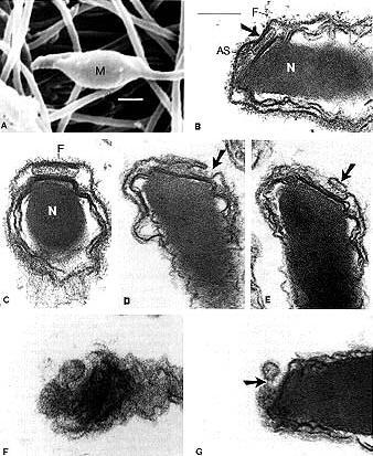

Fig. A Scanning electron microscope image of a Ciona intestinalis spermatozoon. M, mitochondrion. Bar: 1um.
Figs. B,C Sagittal and transverse (at the level of the acrosome) sections, respectively, through the anterior region of the sperm head. An acrosome (arrow) is present. The acrosomal outer membrane and the overlying plasma membrane are in close contact with each other. An electron-dense plate in the acrosome is obvious in B. AS, apical substance; F, fuzzy material; N, nucleus. Bar:200nm (this scale is also applicable to C-G).
Fig. D Longitudinal slightly oblique section through the apex of the sperm head. Membrane fusion between the acrosomal outer membrane at its peripheral margin and the overlying plasma membrane has occurred(arrow).
Fig. E Longitudinal slightly oblique section through the apex of the sperm head. The membrane protrusion made up of the acrosomal outer membrane and the overlying plasma membrane is present(arrow).
Fig. F Longitudinal slightly oblique section through the apex of the sperm head. Fusion between the acrosomal outer membrane and overlying plasma membrane seems to occur along the peripheral margin of the acrosome.
Fig. G Sagittal section through the anterior region of the sperm head. Shedding of a vesicle bounded by fused membrane which is made up of the acrosomal outer membrane and the overlying plasma membrane is probably about to occur. A thin connection between the vesicle and the anterior tip of the sperm head is still present(arrow).


