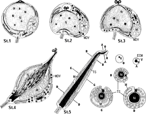

Stage 1: An early spermatid containing a spherical nucleus and a single round mitochondrion. A small blister is present at the apex of the spermatid.
Stage 2: The blister at the anterior tip of spermatid is is enclosed by cytoplasm. In the cytoplasmic lumen, the blister transforms into a dumbbell-shaped protrusion which contains two small vesicles. A well developed Golgi apparatus and vesicles of various sizes which contain electron-dense material are present. In longitudinal sections, the nucleus takes on a heart-shaped apperance.
Stage 3: The dumbbell-shapeds protrusion becomes exposed.
Stage 4: An advanced spermatid with an elongating nucleus in which the chromatin strands are arranged parallel to one another and the mitochonrion elongates antero-posteriorly alongside the nucleus. A well developed Golgi apparatus and vesicles of various sizes are present from stage 2 through stage-4.
Stage 5: A fully differentiated spermatozoon which contains an acrosomal vesicle, an elongated nucleus, tubular sacculi and a single mitochondrion that is moderately wound around the nucleus throughout most its length.
Schematic illustrations of O,A,B,C and D show transverse sections at level O-O, A-A, B-B, C-C and D-D which are indicated on the differentiated spermatozoon. G,Golgi apparatus; MDV, moderately electron-denxe vesicle; M, mitochondrion; TS, tubular sacculus; N, nucleus; ECM, extracellular material; V, acrosomal vesicle.
for details : Bulletin of the College of General Education (Nagoya City Univ.) Vol.38:33-48 (1992)


